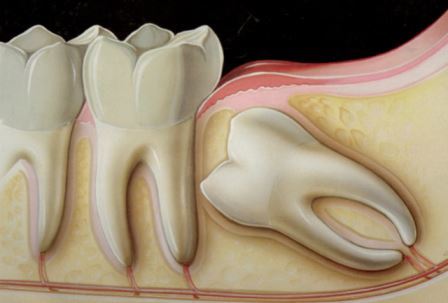
A wisdom tooth may remain partly or fully within the gum or jawbone. Not every impacted tooth must be removed; the decision depends on symptoms, infection risk, damage to the neighbouring tooth, cystic change and its anatomical position.
Clinical examination is combined with panoramic imaging and, when indicated, three-dimensional dental tomography. This helps the surgeon assess the roots and their relationship with the mandibular canal, sinus and adjacent structures.
The procedure is planned under local anaesthesia. We explain the surgical approach, expected recovery and the precautions that matter for your individual case. Swelling and discomfort can vary, and following the post-operative instructions supports healing.
Persistent pain, recurrent swelling, difficulty opening the mouth or an unpleasant taste should be assessed rather than managed only with pain relief.
Every person has different clinical needs. Suitability, treatment options and timing can only be confirmed after an examination and any necessary imaging.