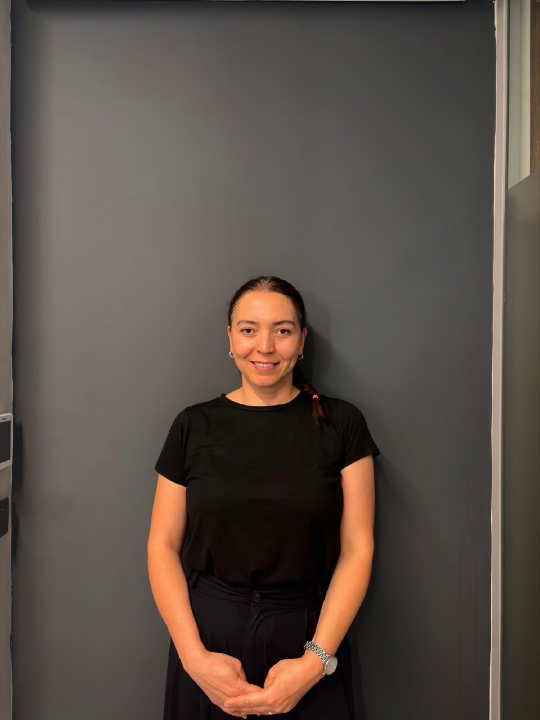
CLINIC COORDINATION
Oya
Clinic Coordinator
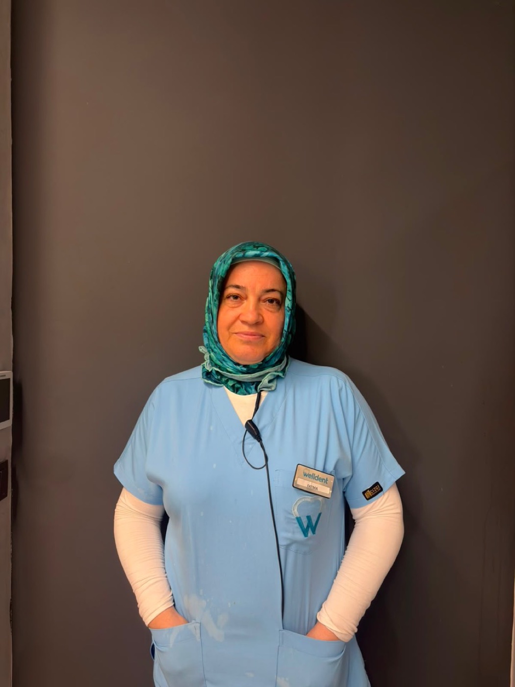
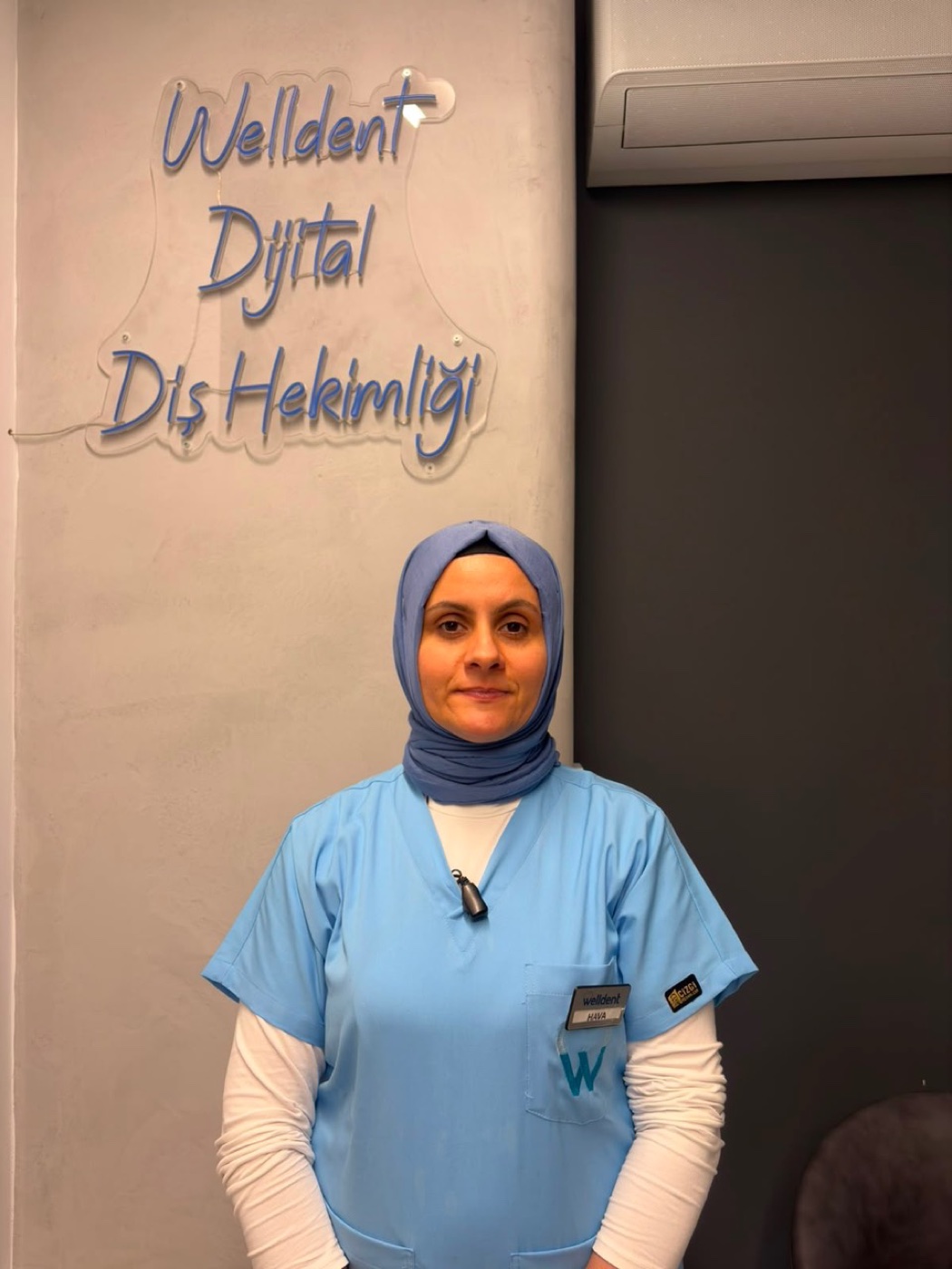
THE WELLDENT TEAM
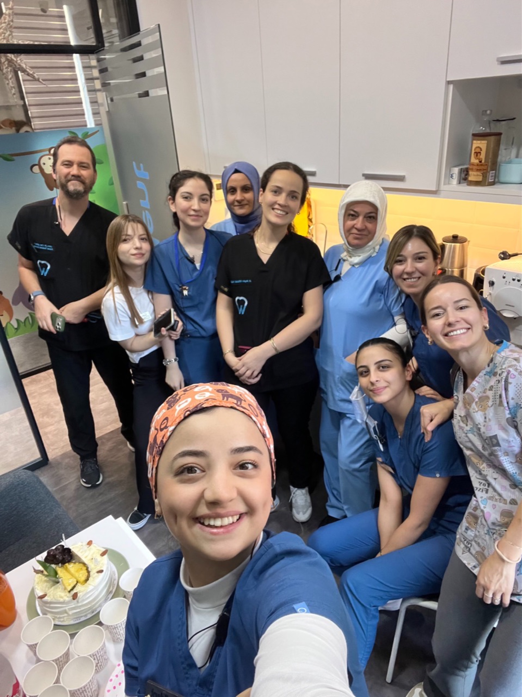
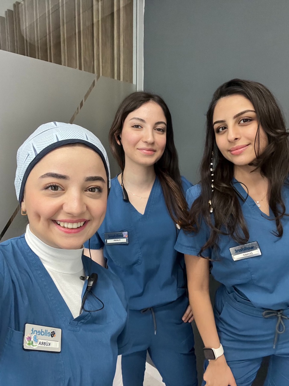
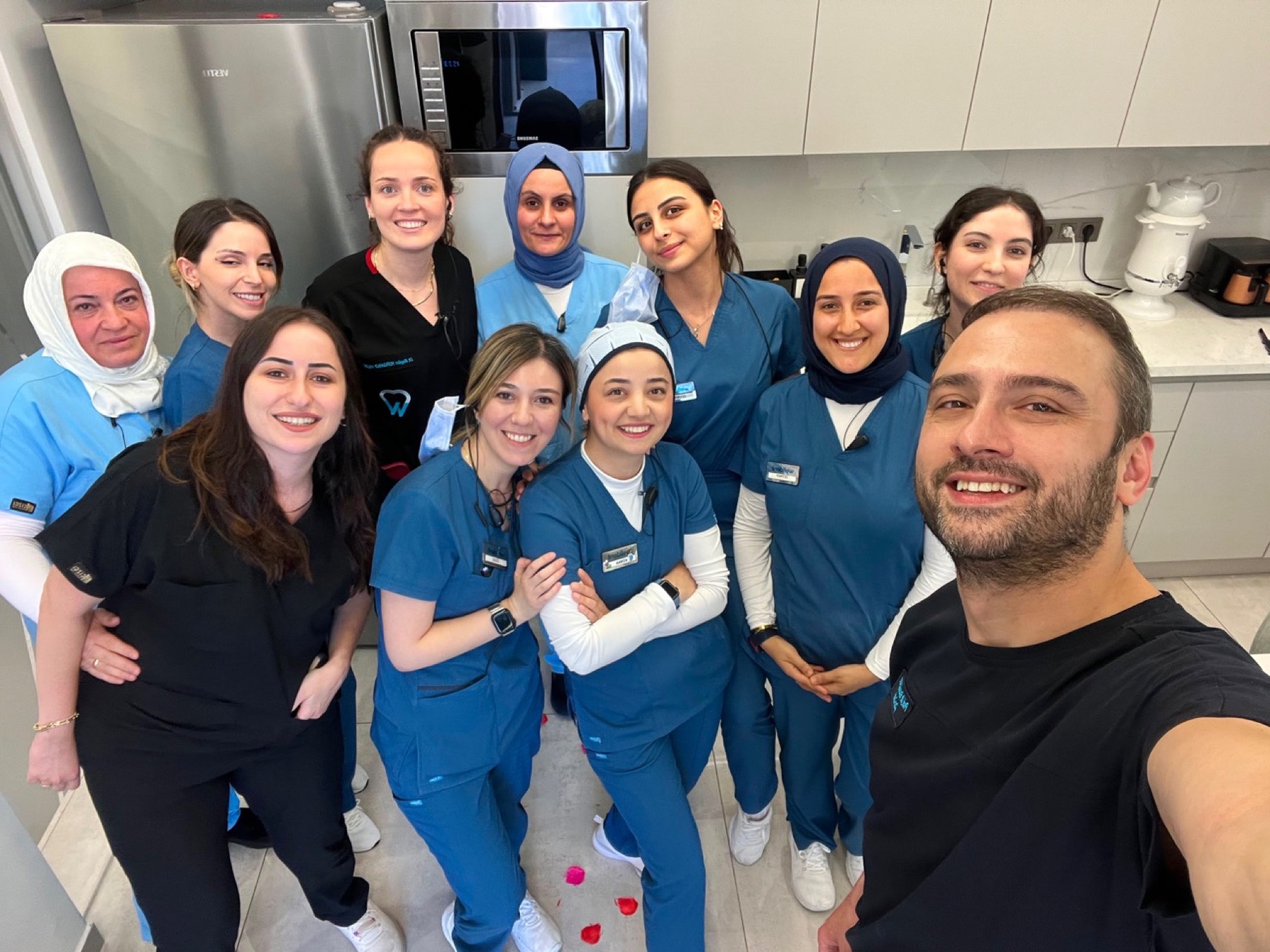
From your first welcome to clinical preparation, hygiene and coordination, every detail is supported by a team working carefully towards the same goal.
WORKING TOWARDS ONE GOAL
Patient reception, clinical preparation, hygiene and administration are all parts of the same experience. By listening to and supporting one another, we aim to keep every visit calm, organised and easy to understand.
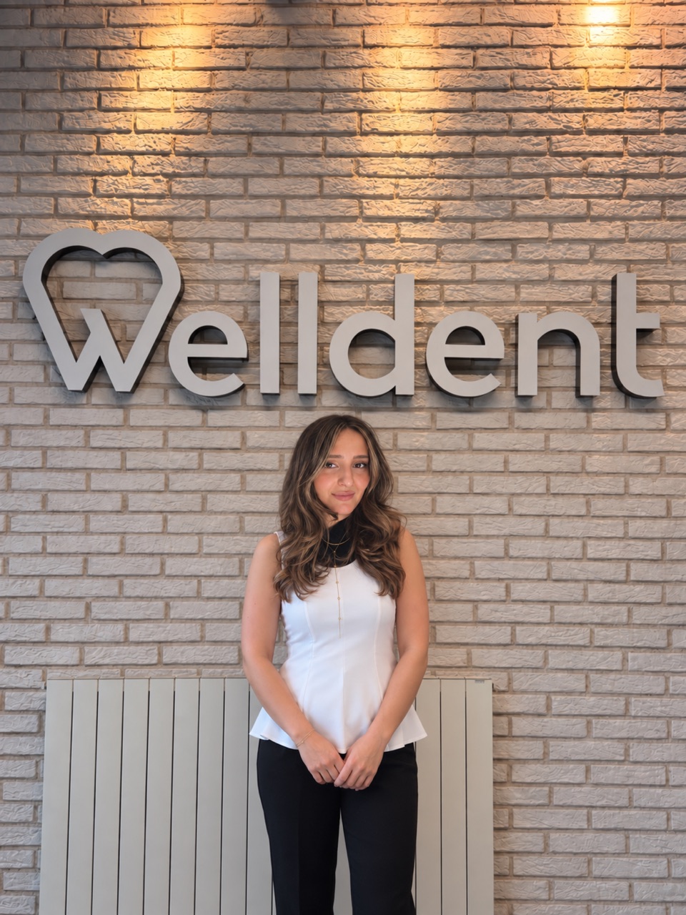
COORDINATION AND RECEPTION
We support open communication and a well-coordinated journey from the first point of contact through the clinic experience.
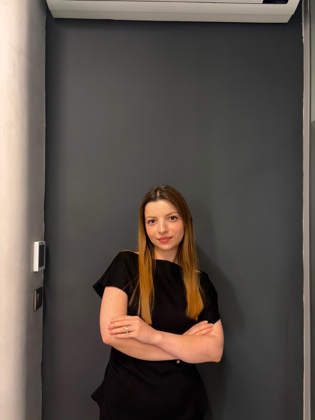
DENTAL ASSISTANT TEAM
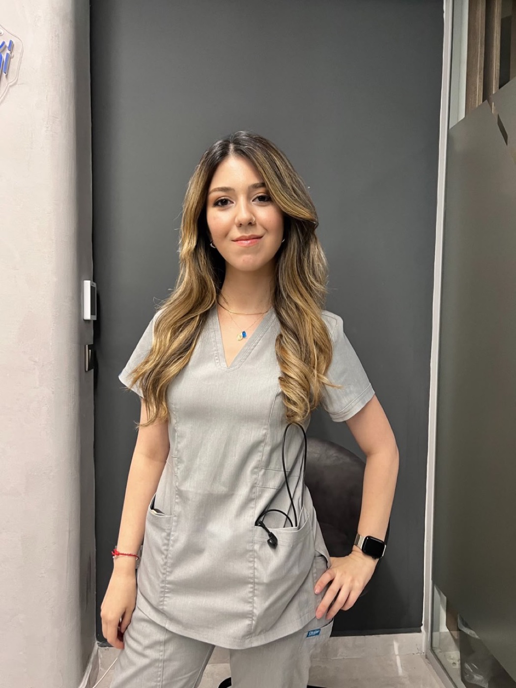
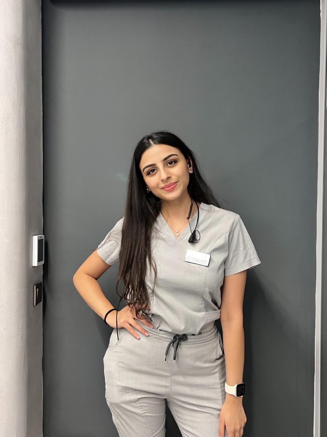
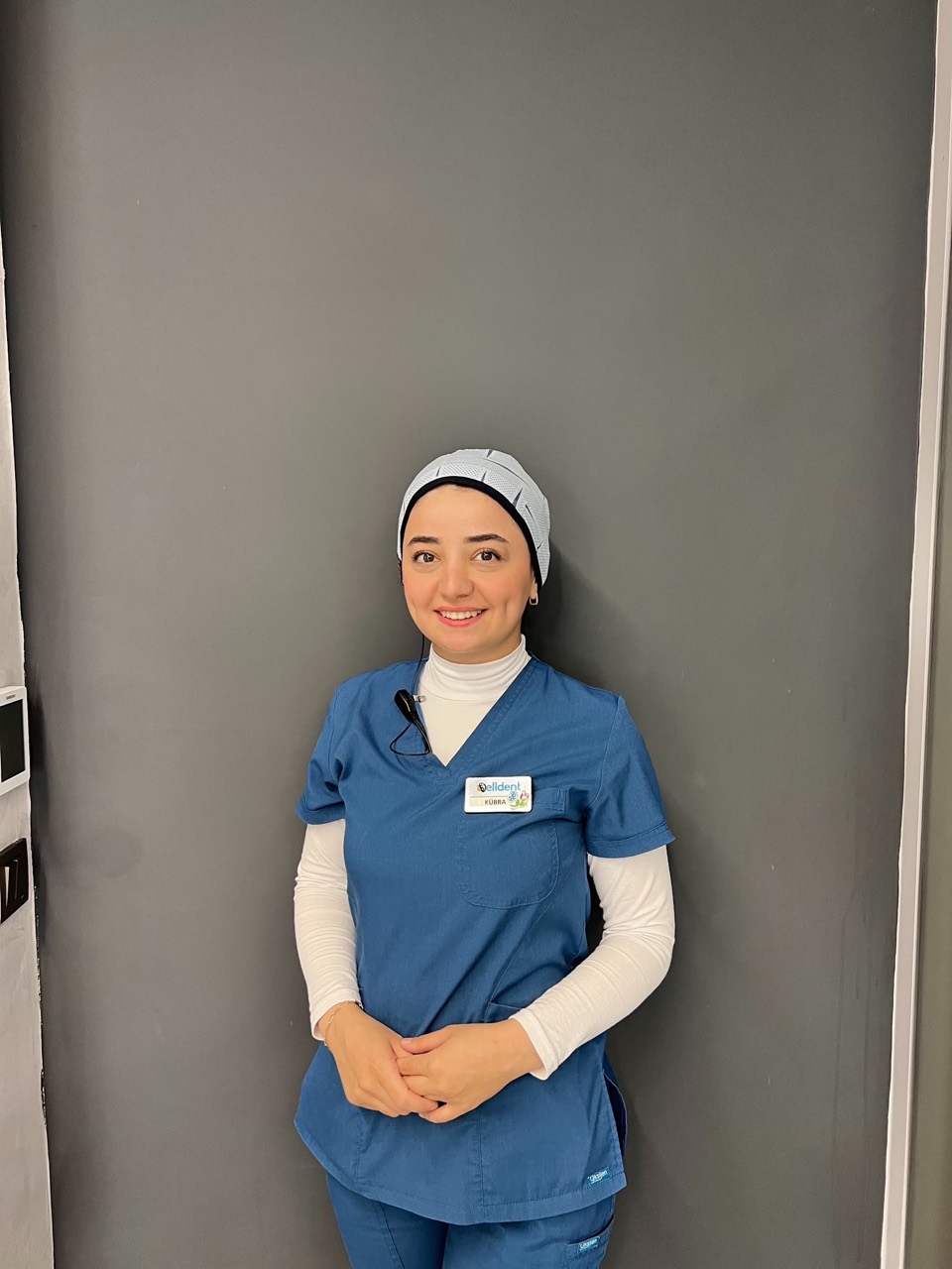
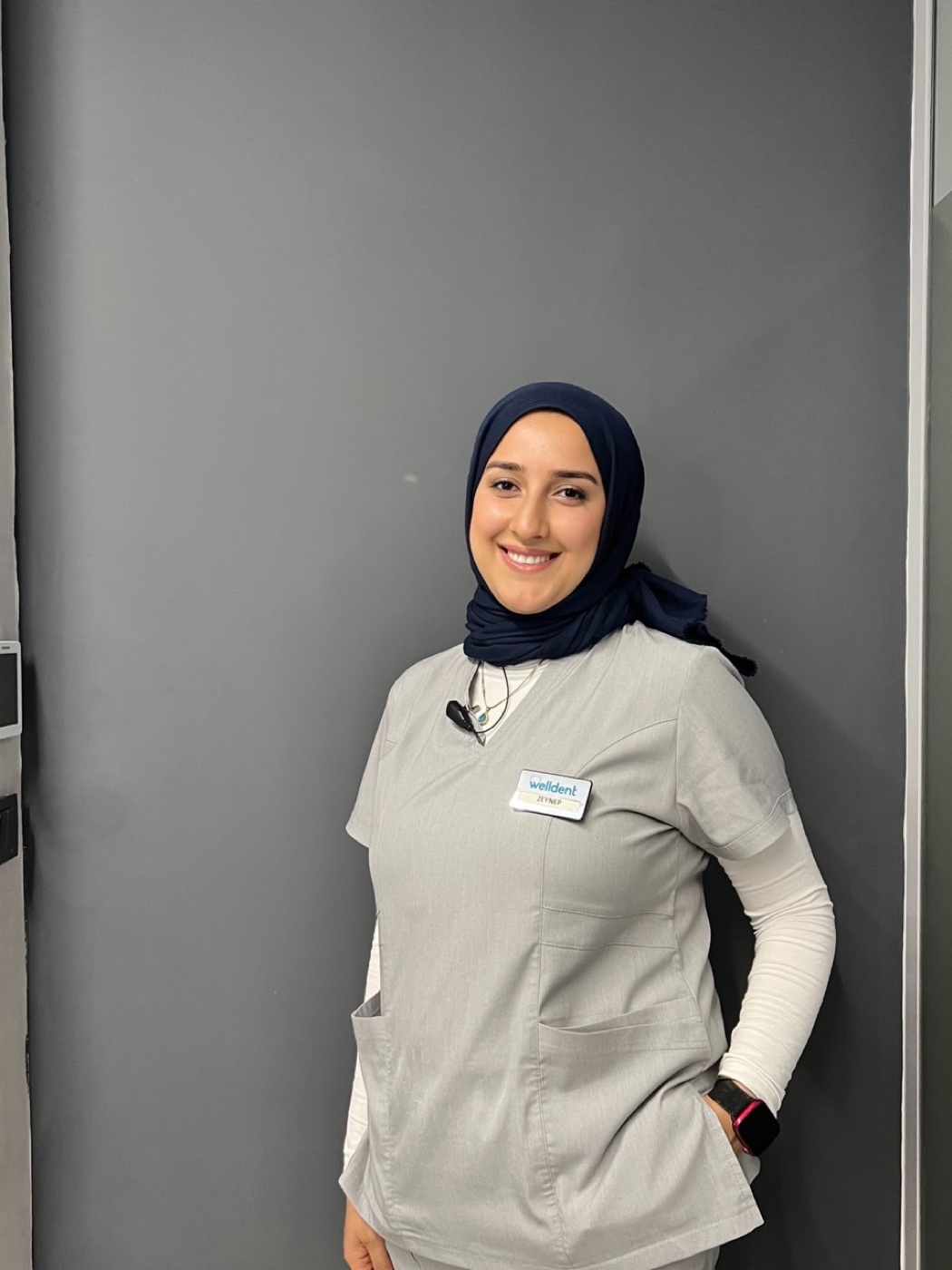
Our assistants work together from treatment-room preparation through chairside clinical support. They follow the cleaning, packaging and sterilisation cycle of reusable dental instruments and store sterile materials under appropriate conditions. Regular material and stock checks help the clinical workflow continue smoothly.
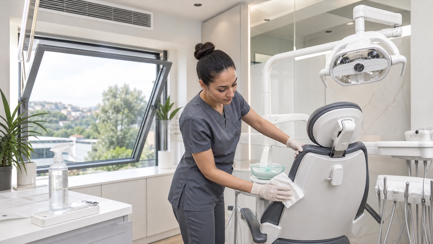
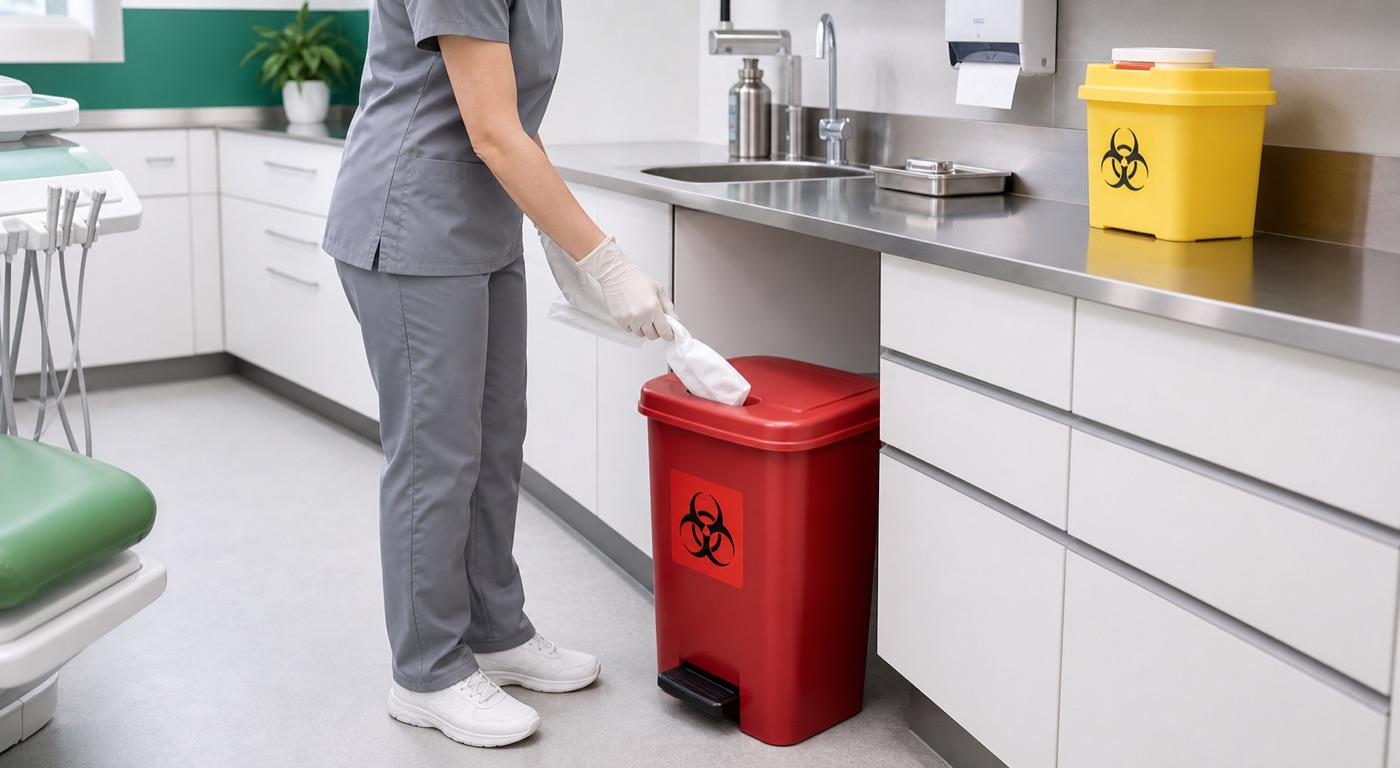
HYGIENE TEAM
ACCOUNTING AND ADMINISTRATION
We help keep accounting and financial processes organised, controlled and aligned with the clinic's internal workflow.

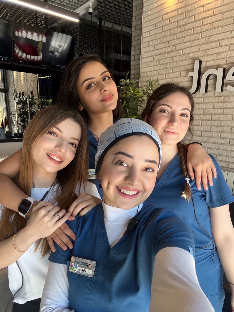
THE STRENGTH OF TEAMWORK
Clear information, careful preparation and coordinated steps help our patients feel understood and supported throughout their visit.
WELLDENT
Our dentists and clinic team are here to listen and guide you.
Book an appointment →DIGITAL DENTISTRY
We use imaging, scanning, magnification and controlled-delivery systems according to individual clinical needs.
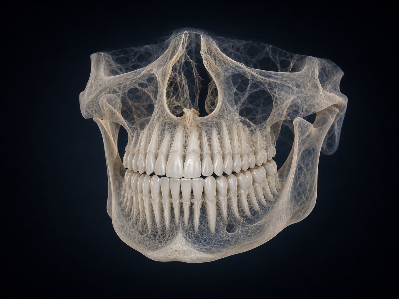
01 · THREE-DIMENSIONAL IMAGING
Cross-sectional and three-dimensional information about teeth, jaws and nearby anatomical structures.
It can support implant planning, impacted-tooth assessment, endodontics and surgical decisions when two-dimensional imaging is not sufficient.
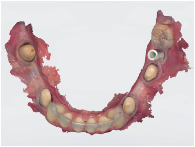
02 · DIGITAL IMPRESSIONS
Optical scanning creates a colour three-dimensional model of teeth and oral tissues.
The scan can support orthodontic, clear-aligner, crown, bridge and implant-restoration workflows without conventional impression material.
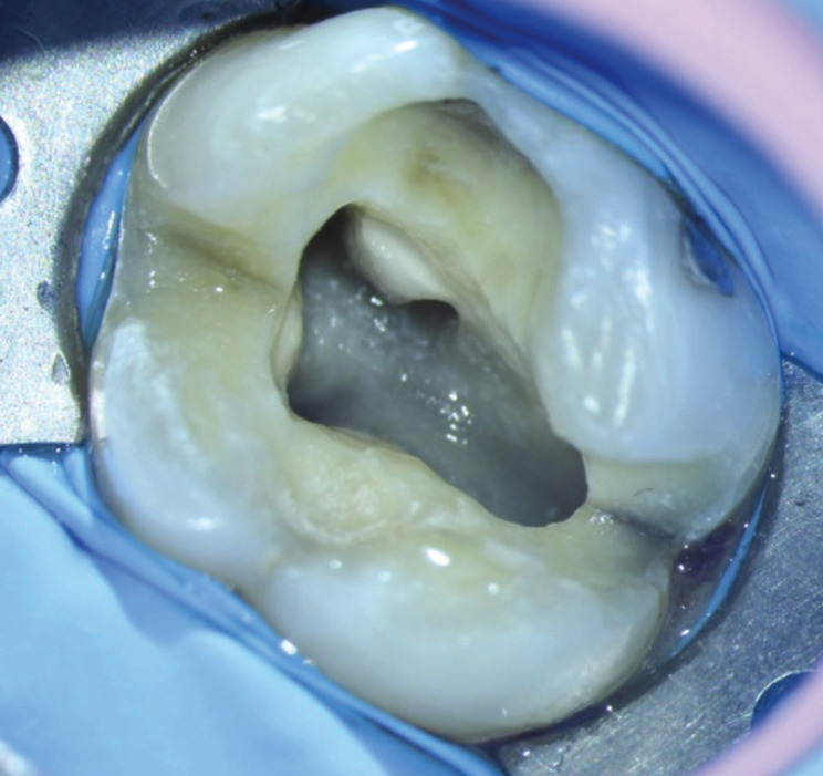
03 · MAGNIFICATION AND LIGHT
Strong magnification and coaxial illumination improve visibility of fine clinical details.
It is particularly useful in endodontics for complex canal anatomy, narrow canal entrances and visually controlled treatment.
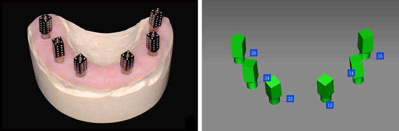
04 · IMPLANT PHOTOGRAMMETRY
A photogrammetry system used to record the three-dimensional position and orientation of implants in selected full-arch cases.
The position data can be combined with an intraoral scan and transferred into a digital restorative workflow.
05 · CONTROLLED LOCAL ANAESTHESIA
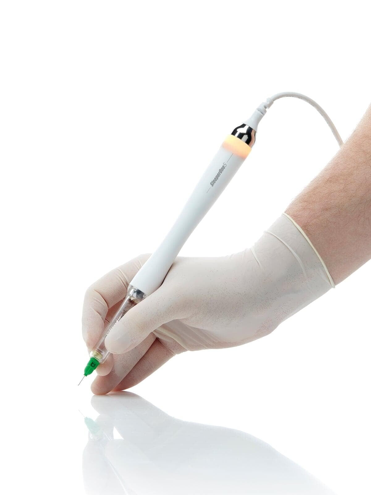
SleeperOne delivers local anaesthetic at a controlled rate through a light, pen-shaped handpiece.
Controlled flow may help reduce the pressure sensation associated with injection. Patients can still feel brief contact or pressure before the familiar numbness develops.
06 · PROFESSIONAL TOOTH WHITENING
Clinical whitening is planned after assessing oral health, starting shade, expectations and the possibility of sensitivity.
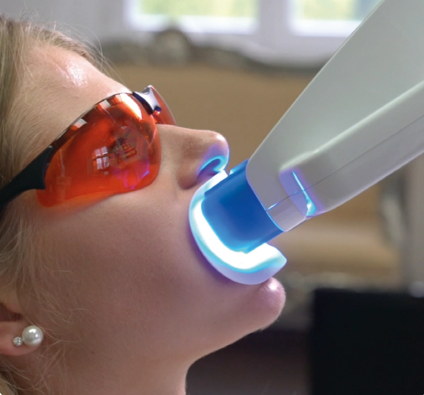
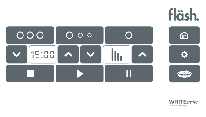
Before treatment, teeth and gums are examined and existing restorations are considered. The lips and soft tissues are isolated, a gingival barrier is applied and protective eyewear is used. The Fläsh system activates the whitening gel with visible blue LED light in timed cycles.
You may notice mild warmth or temporary sensitivity; experience varies from person to person. Natural teeth may lighten, but fillings, crowns and veneers do not change colour. The achievable shade and how long it lasts depend on the starting shade, the cause of discolouration and individual habits.
07 · GUIDED IMPLANT SURGERY
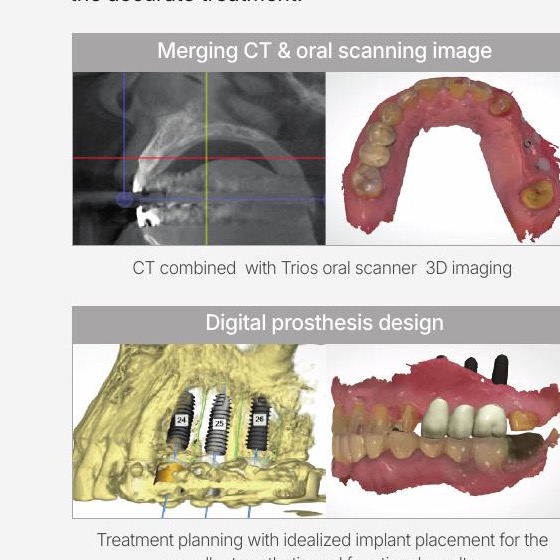
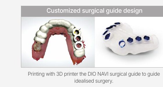
The implant position is planned in advance, and a patient-specific guide helps transfer that plan to the mouth in a controlled way.
CBCT is used to assess bone and nearby anatomy. A 3Shape intraoral scan is combined with this information, allowing the clinician to plan implant position, angle and depth while considering the future teeth.
DIO NAVI is a digital guided-surgery system from DIO Implant, a South Korean company based in Busan. It does not replace clinical judgement; it supports accurate transfer of the clinician-approved plan.
THE DIO NAVI TREATMENT JOURNEY
Every stage is shaped by clinical findings, healing and individual needs. This is the general pathway for suitable full-arch fixed-teeth cases.
We begin with your concerns, expectations and medical history, then assess the remaining teeth, gums and bite as a whole.
CBCT provides detailed information about bone and nearby anatomy. A 3Shape scan records teeth and oral tissues digitally without conventional impression material.
The imaging and scan data are transferred securely to the DIO planning centre. A patient-specific surgical guide is produced from the treatment project approved by the clinician.
When appropriate, necessary extractions and guided implant placement can be completed in the same visit. If adequate initial implant stability is achieved, a screw-retained fixed provisional restoration may be fitted that day.
After approximately three to four months of healing, a clinical review is completed. New imaging and intraoral scans may be taken when needed for the definitive zirconia restoration.
Following digital design, try-in and production, the fit, bite and cleanability of the definitive screw-retained zirconia restoration are checked before delivery, usually over the following one to two months.
Why can timing vary? Bone quality, initial implant stability, healing, additional surgical needs and restorative try-ins can all affect the schedule. Same-day fixed provisional teeth cannot be guaranteed for every patient; suitability is confirmed clinically during surgery.
AN IMPORTANT PRINCIPLE
No device is required for every patient or every treatment. Your clinician will explain which imaging or digital method is relevant to your findings.